Understanding Structure and Function of Muscle Fibers
Let's get a good understanding of all three muscle fibers in our bodies.
The Optimum Workout requires equal parts knowledge, experience and sweat. This month let's work on your knowledge base by delving (deeply, of course) into the way muscle responds to exercise. We start with the structure and function of normal muscle tissue.
Basic Structure
Skeletal muscles vary greatly in size and shape, from the large muscles that control locomotion (gluteus, hamstrings, quadriceps and others) to the tiny muscles of the inner ear. At a more microscopic level, however, skeletal muscles are very similar in structure and function.Muscles are organized in several levels. At the highest level is the whole muscle. The whole muscle is made of many fascicles-the strands of muscle tissue you can see when you look at a piece of roast beef. Each fascicle is made of many muscle fibers. Each fiber, in turn, is made of 500 to 10,000 myofibrils. Each myofibril is made of 1,000 to 2,000,000 sarcomeres laid end-to-end. Each sarcomere is made of overlapping thick and thin filaments, which are made of contractile and regulatory proteins-actin, myosin, tropomyosin, troponin and others. The contractile and regulatory proteins are the foundation of muscle contraction.
Voluntary muscle contraction requires a number of steps. It starts with an electrical signal in the brain, a signal to contract. The signal travels along a nerve, first to the spinal column, then to the muscle. When the nerve approaches the muscle, the nerve branches into many tiny fibers, which extend throughout the muscle. The electrical signal travels along the nerve fibers and essentially reaches all parts of the muscle at the same time.
At the end of each nerve fiber is a place where the nerve actually meets the muscle fibers, called the neuromuscular junction. The electrical signal crosses the neuromuscular junction and is transmitted deep inside the muscle fibers.
Here the signal stimulates the release of calcium inside the muscle, which causes the thick and thin filaments to slide across one another. When the filaments slide in this way, the sarcomere shortens, generating force. The activity of billions of sarcomeres shortening in a muscle all at once results in the contraction of the whole muscle.
The energy that causes the thick and thin filaments to slide across one another comes from mitochondria, the "factories" inside muscle cells that generate energy from glucose. Inside the mitochondria are enzymes that control the chemical reactions involved in breaking down glucose. Both resistance and strength training cause changes in mitochondria and mitochondrial enzymes.
Muscle Fiber Types
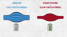
Not all muscle fibers are created equal. In humans there are three main types of fibers: 1, 2A and 2B. Type 1 fibers are slow to contract but slow to fatigue. Type 2A are fast to contract and intermediate to fatigue. Type 2B are fast to contract and fast to fatigue.One of the reasons Type 1 fibers are slow to fatigue is that they have more mitochondria, which makes them capable of producing more energy. This decreases fatigability. Type 1 fibers also are smaller in diameter than Type 2A and 2B fibers and have increased capillary blood flow around them. Having a smaller diameter and increased blood flow improves oxygen delivery and waste-product removal from muscle fibers, which also decreases fatigability.
All muscles contain all three fiber types but in differing amounts. The postural muscles of the back, for instance, have to be contracted much of the time to keep your upper body upright. These muscles have a high percentage of Type 1 (slow) fibers, so they fatigue slowly and recover quickly.
The biceps, on the other hand, are at rest most of the time. When called into action, they usually have to perform brief bouts of intense activity- for example, a set of biceps curls. Your biceps tend to fatigue quickly and recover slowly because they have a high percentage of Type 2 (fast) fibers.
When a muscle begins to contract, primarily Type 1 fibers are activated first, then Type 2A, then 2B. This sequence of fiber recruitment allows very delicate and finely tuned muscle responses to brain commands. It also makes Type 2B fibers difficult to train; most of the Type 1 and 2A fibers have to be activated before a large percentage of the 2B fibers participate.
One way to remember the differences between muscle with predominantly Type 1 (slow) fibers and muscle with predominantly Type 2 (fast) fibers is to think chicken.
Everyone is familiar with dark meat and white meat. Chicken legs- dark meat-have to hold up the chicken most of the day. Not surprisingly, the chicken leg consists of muscle tissue that is slow to fatigue and fast to recover. This corresponds to muscle with predominantly Type 1 fibers in humans. Dark meat is dark because it has a greater number of mitochondria (mitochondria are dark). Having more mitochondria gives chicken legs a greater capacity to make energy, making them slow to fatigue and fast to recover.
On the other hand, chicken breasts-white meat-are at rest most of the time but are called on for brief bouts of intense activity, for example, a few seconds of flying around the barnyard. Chicken breast consists of muscle tissue that can contract quickly but is fast to fatigue and slow to recover. Breast meat is whiter in color because it has fewer mitochondria.
The darkest meat of all is heart muscle, which can never afford to tire. Heart muscle cells are more than 50 percent mitochondria by volume.
In addition to more mitochondria, dark meat has greater fat stores in its muscle fibers, and these serve as a source of energy for the mitochondria. Result? The extra fat in dark meat makes it moister but higher in calories than white meat.